Using theoretical calculations of forces and currents, researchers in Spain and the Czech Republic have shown that the bright spots seen in scanning-tunnelling and atomic-force microscope images may correspond to the spaces between atoms and not to the atoms themselves. The result could help answer a decades-old question of whether such microscopy techniques actually image atoms or not.
Nanotechnology as a subject was born in the 1980s with the advent of microscopes capable of imaging materials on the atomic scale. For example, the development of the scanning tunnelling microscope (STM) allowed the distances between individual atoms to be resolved for the first time. An STM involves placing a tiny metal tip very near to the surface of interest and applying a voltage between the surface and tip. The tip is then scanned just above the surface and an image generated by measuring the current of electrons that tunnel between the tip and surface.
The images produced are actually spatial variations in the electron density of states of the surface near the Fermi level – the energy level of the most loosely held electrons in a solid. However, since the density of states does not always peak when the tip is directly above the atoms, it is not possible to know with absolute certainty whether the maxima (or bright spots) in STM images correspond to atoms or the hollow spaces between atoms. What is more, images of the same surface can appear completely different depending on the structure and composition of the STM tip, so interpreting the images obtained means that researchers need to understand the short-range chemical forces between this tip and a surface.
Perfect test beds
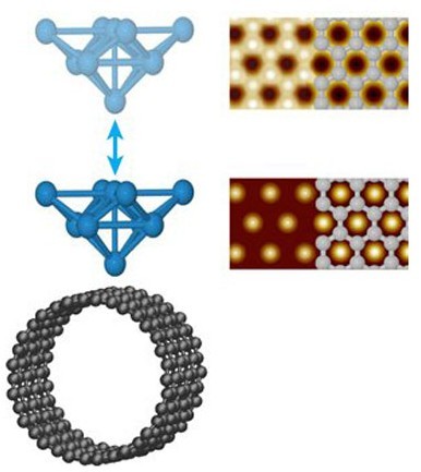
Rubén Pérez and colleagues at the Universidad Autonóma de Madrid and the Czech Academy of Sciences have now carried out an extensive set of first-principles calculations to map out the interaction between the tip and samples of single-wall carbon nanotubes and graphite. The unique mechanical and electronic properties of carbon-based materials such as fullerenes, nanotubes, graphene and nanoribbons makes them extremely promising for a wide range of technology applications and their simple honeycombed structure makes them perfect test beds for STM imaging.
Pérez and co-workers' simulations combine density functional theory (DFT) calculations for the short-range chemical force between the tip and sample combined with semi-empirical atomistic approaches for the longer-range Van der Waal's interaction between the two objects. "This methodology is particularly needed in the case where experiments seems to suggest that the Van der Waal's interaction competes, and even dominates, over the short-range force in the distance range relevant for STM imaging," explains Pérez.
The results show that in the "near-contact" regime, bright spots in the final STM images correspond to hollow positions between atoms rather than atomic sites themselves.
Better understanding of defects
According to the researchers, the findings will help them understand the fundamental mechanisms behind STM imaging and so better characterize defects in carbon nanostructures. Such defects, which include single-atom vacancies and dopants in graphene and carbon nanotubes, play a crucial role in the electronic and magnetic proprieties of these materials. The results should also help guide scientists in their choice of tip for stable, high-resolution STM imaging because different tips interact to a greater or lesser extent with the sample surface.
The team is now applying its methods to explore the electronic properties of epitaxial graphene on metals. "These calculations will also shed light on the possible magnetic state associated with defects in graphene and graphite – something that has been predicted in theory but not yet experimentally confirmed," Pérez told physicsworld.com.
'Excellent result'
"This is an excellent result from some of the foremost scanning-probe theorists in the world," commented Philip Moriarty of the University of Nottingham in the UK, who was not involved in the work. "Scanning-probe-microscope images are notoriously difficult to interpret because the probe itself plays an integral role in the image-formation process. All scanning-probe microscopists working on atomic-resolution imaging (and manipulation) of surfaces and nanostructures will now be able to benefit from this work."
The results were published in Phys. Rev. Lett. 106 176101.
Source: nanotechweb.org